目录图表
谷胱甘肽(GSH)几乎存在于所有人体细胞中,在控制细胞氧化还原状态中起着至关重要的作用[1–5]。GSH浓度异常与许多疾病密切相关,如艾滋病、癌症、肝损伤和衰老引起的疾病[2]。因此其检测方法层出不穷,如电致发光[6]、电化学法[7]、高效液相色谱(HPLC)[8]、毛细管电泳[9]、质谱(MS)[10]、表面增强拉曼散射(SERS)[11]、荧光法[12]等,这些方法因为复杂的操作程序,对专业检测人员要求较高,而且操作过程耗时、仪器昂贵、灵敏度不高、专一性差。比色法相较于其他方法,由于其快速、成本低和可见性好等优点引起了广泛关注。
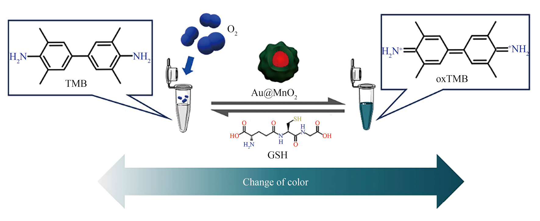
模拟酶3,3′,5,5′-四甲基联苯胺(TMB)和O2体系是比色检测GSH最常用的方法之一。在包括碳基纳米材料[13–15]、无机纳米颗粒[16–17]和金属有机骨架基纳米材料[18–19]等模拟酶的催化下,TMB被O2氧化后,溶液颜色从无色变为蓝色。加入GSH后,蓝色逐渐褪去。基于此,本文制备了一种核壳纳米结构的Au@MnO2复合材料,这种Au@MnO2复合材料在氧气存在下,无需添加额外的氧化剂即可显示出较强的催化活性,将TMB氧化成TMB的氧化产物(oxTMB),从而使其溶液的颜色从无色变为蓝色;加入GSH后,TMB的氧化产物(oxTMB)和GSH反应又重新生成了TMB,最终导致溶液蓝色褪去,并伴随着溶液的吸光度变化,因此可以利用该变化比色检测GSH的含量,其检测原理如图1所示。
实验使用试剂有:氯金酸、柠檬酸钠、高锰酸钾(KMnO4)和聚烯丙胺盐酸盐(PAH)购于美国Sigma Aldrich公司。3,3′,5,5′-四甲基联苯胺(TMB)、抗坏血酸(AA)、半胱氨酸(Cys)、茶多酚(TP)、多巴胺(DA)、尿酸(UA)、柠檬酸(CA)、葡萄糖(Glu)、褪黑素(Mel)和谷胱甘肽(GSH)购于上海阿拉丁公司。所有试剂均为分析级,无需进一步纯化即可使用。
测试仪器有:UV-vis 2700型紫外可见分光光度计(日本岛津公司)、H-7560型透射电子显微镜(TEM,日本日立公司)、JSM-7800型扫描电子显微镜(SEM,日本日立公司)和实验级超纯水器(Aquaplore 3S,美国艾科浦公司)。
在剧烈搅拌下将0.74 ml 0.1 mol·L−1 HAuCl4溶液加入295 ml沸腾超纯水中。然后,加入4.5 ml 10 mg·ml−1柠檬酸钠,并加热约15 min,所得溶液冷却至室温并在4 ℃储存。向所得纳米金溶液中添加1.5 ml 22 mg·ml−1 KMnO4溶液。反应5 min后,将3 ml 25 mg·ml−1的PAH滴入溶液中搅拌1 h,直到溶液颜色从紫色变为深绿色。然后将Au@MnO2在10000 r·min−1下离心20 min并用纯水洗涤3次后获得Au@MnO2,净化后的Au@MnO2分散在去离子水中以备将来使用[20]。
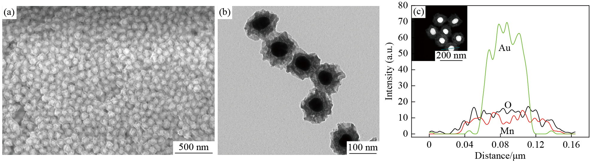
图2为Au@MnO2纳米粒子的SEM图像、TEM图像和线性扫描图。可见,所制备的Au@MnO2纳米粒子具有类似于飞碟状的纳米结构,其中,纳米核是平均尺寸约为50 nm的金纳米粒子,壳是厚度为20~30 nm的MnO2,产生了均匀的核壳结构(2(b))。图2(c)的线性扫描图表明Au@MnO2纳米粒子由Au,Mn和O元素组成。
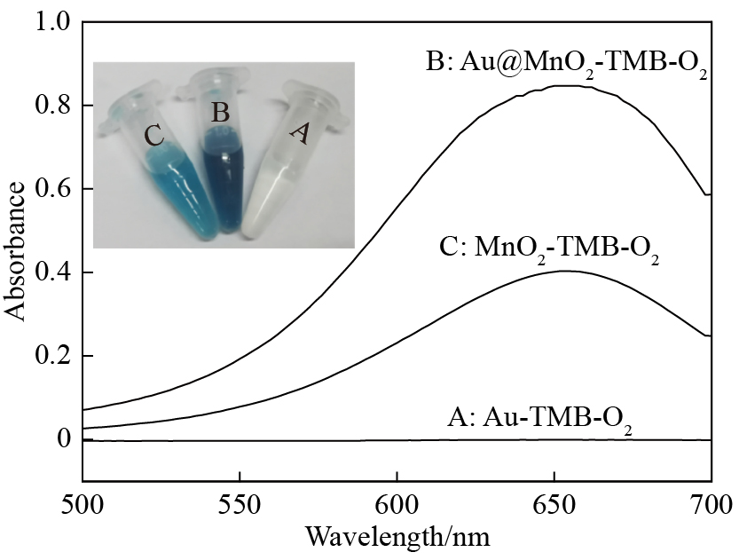
通过UV-vis研究了Au,MnO2和Au@MnO2的氧化物酶催化活性,如图3所示。可见,MnO2和Au@MnO2在O2存在下催化TMB的氧化,在650 nm处有一个明显的UV-vis吸收峰,溶液颜色为蓝色。与MnO2-TMB-O2体系相比,Au@MnO2-TMB-O2体系表现出更强的吸收峰和更蓝的溶液颜色,表明Au@MnO2比MnO2具有更好的氧化物酶性能。对于Au-TMB-O2系统,其在650 nm处的吸收峰几乎可以忽略不计。
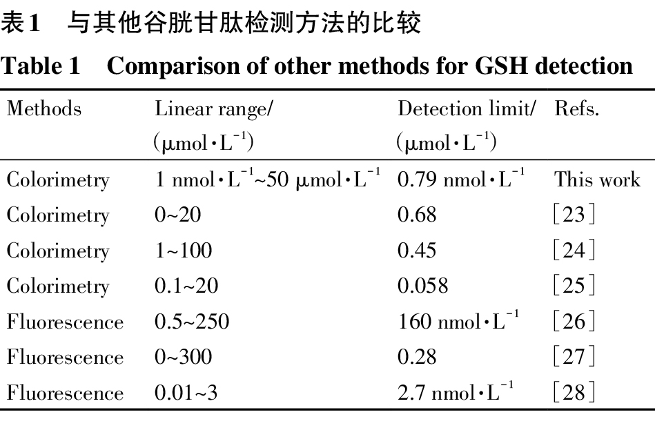
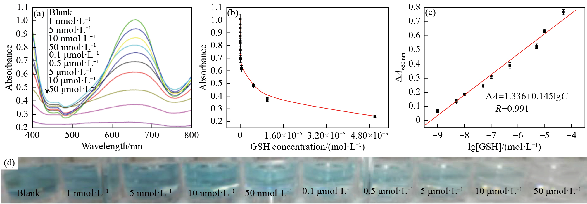
图4为含有不同浓度GSH的oxTMB溶液的UV-vis吸收光谱,溶液吸光度和谷胱甘肽浓度与GSH浓度对数之间的关系以及相应溶液颜色照片。可见,在最优条件(pH为6,MnO2和TMB的反应时间为10 min,GSH的孵化时间为5 min)下,随着GSH浓度从0到5 μmol·L−1的增加,oxTMB在650 nm波长处的吸光度逐渐降低(图4(a,b)),伴随着颜色从深蓝色变为浅蓝色(图4(d))。同时,由图4(c)可知,吸光度变化(ΔA650 nm)与GSH浓度(1 nmol·L−1~50 μmol·L−1)的对数成正比(R=0.991),其中ΔA是不含GSH和含GSH溶液吸光度的差值。基于信噪比(signal-to-noise ratio,S/N)=3[21],检测限(LOD)达到0.79 nmol·L−1,远低于血清中GSH的最低水平(0.5~10 mmol·L−1)[22]。与表1中的其他GSH检测方法相比,该方法对GSH检测更敏感,LOD更低。
| Methods | Linear range/(μmol·L−1) | Detection limit/(μmol·L−1) | Refs. |
|---|---|---|---|
| Colorimetry | 1 nmol·L−1~50 μmol·L−1 | 0.79 nmol·L−1 | This work |
| Colorimetry | 0~20 | 0.68 | [23] |
| Colorimetry | 1~100 | 0.45 | [24] |
| Colorimetry | 0.1~20 | 0.058 | [25] |
| Fluorescence | 0.5~250 | 160 nmol·L−1 | [26] |
| Fluorescence | 0~300 | 0.28 | [27] |
| Fluorescence | 0.01~3 | 2.7 nmol·L−1 | [28] |
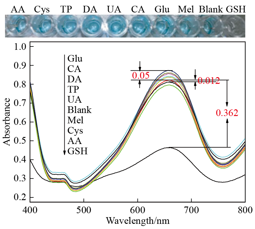
为了考察该传感器对GSH的选择性,采用AA,Cys,TP,DA,UA,CA,Glu和Mel作为干扰物,结果如图5所示(GSH浓度:0.5 μmol·L−1,干扰抗氧化剂浓度:50 μmol·L−1)。可见,这些浓度是GSH浓度100倍的干扰物质对oxTMB吸光度的影响非常小。与空白溶液吸光度相比,这些干扰物引起的吸光度差值在0.012~0.05之间。然而,与空白相比,0.5 μmol·L−1 GSH引起吸光度发生巨大变化,吸光度差值为0.362。以上结果表明,AA,Cys,TP,DA,UA,CA,Glu和Mel对GSH检测几乎没有影响,并且GSH的检测选择性是可以接受的。
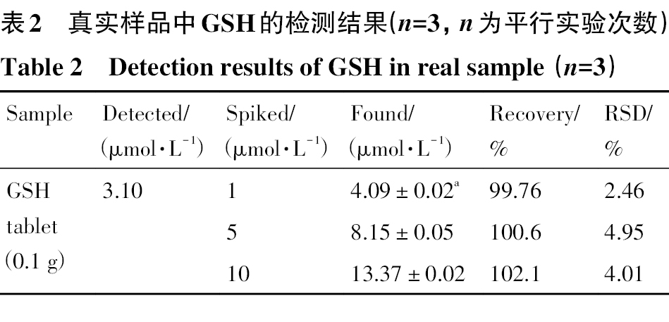
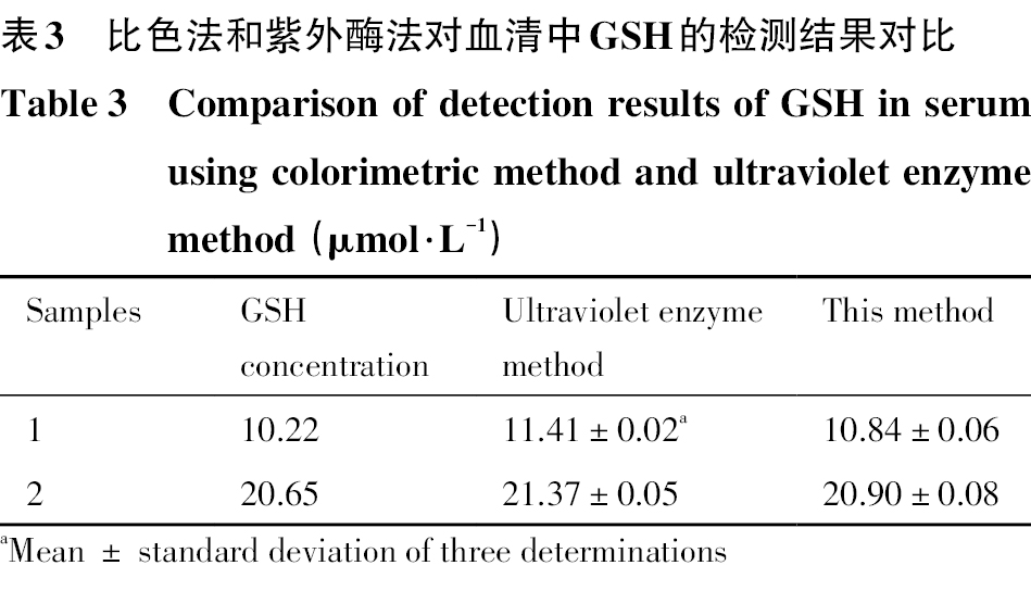
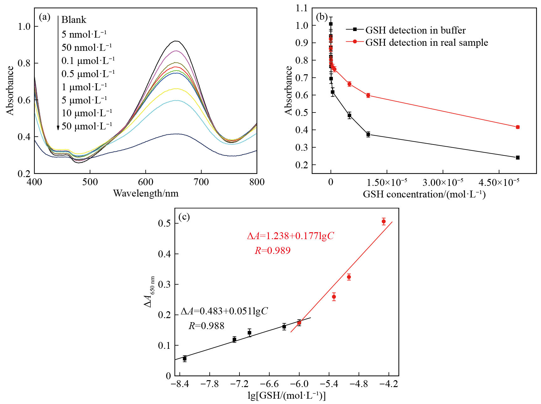
为了评估该方法在复杂真实样品中的可行性,对人血清中的GSH进行了测定。人体血液中GSH的正常含量为26~34 mg·(100 g)−1。为了研究这种传感器对血清样品中GSH的定量检测能力,首先将血清通过硝酸纤维素膜(0.22 μm)过滤以去除杂质,然后将血清稀释100倍,在稀释后的血清样品中加入一定含量的GSH,使血清中GSH的最终浓度在5 nmol·L−1~50 μmol·L−1之间,如图6(a)所示。与缓冲液中GSH的检测结果相比,血清中GSH(0~1 μmol·L−1)的低浓度几乎没有引起吸光率的变化;当GSH浓度超过1 μmol·L−1,出现更高的吸光率差(˂0.223)(图6(b)),这意味着受血清影响的高浓度GSH检测更为严重。然而,鉴于在5 nmol·L−1~1 μmol·L−1和1~50 μmol·L−1范围内,吸光度变化与GSH浓度之间具有良好的线性关系,如图6(c)所示,该传感器适用于实际样品中GSH的定量分析。此外,还使用GSH片剂进行回收试验。如表2所示,GSH片剂(0.1 g)样品的平均回收率为99.76%~102.1%。另外,也将本文的方法和医院常用的紫外酶法检测GSH做了对比。如表3所示,该方法与紫外酶在测定血清中的GSH水平方面具有可比性,表明该方法对实际样品中GSH的检测是可靠的。
| Samples | GSH concentration | Ultraviolet enzyme method | This method |
|---|---|---|---|
| 1 | 10.22 | 11.41±0.02a | 10.84±0.06 |
| 2 | 20.65 | 21.37±0.05 | 20.90±0.08 |
1)通过氯金酸和柠檬酸钠还原法制得纳米金,然后向其中加入KMnO4和PAH溶液成功制备出飞碟状核壳Au@MnO2纳米粒子。该纳米粒子具有很好的类氧化酶性质,能催化TMB和空气中的氧气发生反应,生成蓝色的oxTMB。而GSH的存在抑制了TMB和氧气的反应,使溶液的蓝色变浅。根据这一性质,成功地设计出了基于Au@MnO2-TMB-O2的比色传感策略的高灵敏检测GSH的方法。
2)Au@MnO2纳米粒子基于的传感系统对GSH出示了出色的检测能力,溶液的吸光度变化与GSH浓度在1 nmol·L−1~50 μmol·L−1的浓度范围内呈良好的线性关系,检测限达到0.79 nmol·L−1。在其他的抗氧化剂作为干扰物的情况下,该传感系统对GSH显示出出色的特异性。重要的是,该方法在定量检测人血清中GSH时表现出良好的性能,在医学诊断中显示出潜在的应用前景。
Zhao N,Gong Q,Zhang R X,Yang J,Huang Z Y,Li N,Tang B Z. A fluorescent probe with aggregation-induced emission characteristics for distinguishing homocysteine over cysteine and glutathione[J].Journal of Materials Chemistry A,2015,3(32):8397.
Yin J,Kwon Y,Kim D,Lee D,Kim G,Hu Y,Ryu J H,Yoon J. Preparation of a cyanine-based fluorescent probe for highly selective detection of glutathione and its use in living cells and tissues of mice[J].Nature Protocols,2015,10(11):1742.
Benhar M,Shytaj I L,Stamler J S,Savarino A J. Dual targeting of the thioredoxin and glutathione systems in cancer and HIV[J].Journal of Clinical Investigation,2016,126(5):1630.
Yang P,Ebbert J O,Sun Z,Weinshilboum R M. Role of the glutathione metabolic pathway in lung cancer treatment and prognosis:a review[J].Journal of Clinical Oncology,2006,24(11):1761.
Huang M,Wang H,He D,Jiang P,Zhang Y. Ultrafine and monodispersed iridium nanoparticles supported on nitrogen-functionalized carbon:an efficient oxidase mimic for glutathione colorimetric detection[J].Chemical Communications,2019,55(25):3634.
Chen J,He Z Y,Liu H,Cha C S. Electrochemical determination of reduced glutathione (GSH) by applying the powder microelectrode technique[J].Journal of Electroanalytical Chemistry,2006,588(2):324.
Chen W,Zhao Y,Seefeldt T,Guan X. Determination of thiols and disulfides via HPLC quantification of 5-thio-2-nitrobenzoic acid[J].Journal of pharmaceutical and biomedical analysis,2008,48(5):1375.
Zinellu A,Sotgia S,Scanu B,Usai M F,Fois A G,Spada V,Deledda A,Deiana L,Pirina P,Carru C. Simultaneous detection of N-acetyl-l-cysteine and physiological low molecular mass thiols in plasma by capillary electrophoresis[J].Amino Acids,2009,37(2):395.
Burford N,Eelman M D,Mahony D E,Morash M. Definitive identification of cysteine and glutathione complexes of bismuth by mass spectrometry:assessing the biochemical fate of bismuth pharmaceutical agents[J].Chemical Communications,2003,(1):146.
Li Y X,Li P,Chen Y Q,Wu Y,Wei J.Interfacial deposition of Ag nanozyme on metal-polyphenol nanosphere for SERS detection of cellular glutathione[J].Biosensors & Bioelectronics,2023,228:115200.
Yang J J,Bian L,Wang A Q,Zhao X F,Arslanov T,Zeng L,Mu B,Song M X,Liu P,Gu X B. Interfacial interactions in CdTe/MX (M=Zn,Cd,Sb,Bi;X=Te,Se,S,O) nanoparticles:implications for glutathione fluorescence detection[J].ACS Applied Nano Materials,2025,8(35):17031.
Qi H,Teng M,Liu M,Liu S,Li J,Yu H,Teng C,Huang Z,Liu H,Shao Q. Biomass-derived nitrogen-doped carbon quantum dots:highly selective fluorescent probe for detecting Fe3+ ions and tetracyclines[J].Journal of Colloid and Interface Science,2019,539:332.
Cai N,Tan L,Li Y,Xia T T,Hu T Y,Su X G. Biosensing platform for the detection of uric acid based on graphene quantum dots and G-quadruplex/hemin DNAzyme[J].Analytica Chimica Acta,2017,965:96.
Liu Q Y,Yang Y T,Li H,Zhu R,Shao Q,Yang S G,Xu J J. NiO nanoparticles modified with 5,10,15,20-tetrakis (4-carboxyl pheyl)-porphyrin:promising peroxidase mimetics for H2O2 and glucose detection[J]. Biosensors & Bioelectronics,2015,64:147.
Liu Q Y,Yang Y T,Lv X T,Ding Y N,Zhang Y Z,Jing J J,Xu C X. One-step synthesis of uniform nanoparticles of porphyrin functionalized ceria with promising peroxidase mimetics for H2O2 and glucose colorimetric detection[J].Sensors and Actuators B:Chemical,2017,240:726.
Zhang X L,Li G L,Wu D,Li X L,Hu N,Chen J,Chen G,Wu Y N. Recent progress in the design fabrication of metal-organic frameworks-based nanozymes and their applications to sensing and cancer therapy[J]. Biosensors & Bioelectronics,2019,137:178.
Wang X,Feng S Y,He D P,Jiang P.Porous manganese-cobalt oxide microspheres with tunable oxidase mimicking activity for sulfide ion colorimetric detection[J].Chemical Communications,2020,56(90):14098.
Casado A G,Rodriguez L C,Hernandez E A,Vilchez J L. Estimate of gas chromatographic blanks-application to detection limits evaluation as recommended by IUPAC[J]. Journal of Chromatography A,1996,726(1-2):133.
Li T,Wang Z,Jiang D,Wang H,Lai W F,Lv Y,Zhai Y. A FRET biosensor based on MnO2 nanosphere/copper nanocluster complex:from photoluminescence quenching to recovery and magnification[J].Sensors and Actuators B-Chemical,2019,290:535.
Liu Y F,Zhou M,Cao W,Wang X Y,Wang Q,Li S R,Wei H. Light-responsive metal-organic framework as an oxidase mimic for cellular glutathione detection[J].Analytical Chemistry,2019,91(13):8170.
Jin C Q,Lian J J,Gao Y,Guo K K,Wu K L,Gao L N,Zhang X X,Zhang X,Liu Q Y. Si doped CoO nanorods as peroxidase mimics for colorimetric sensing of reduced glutathione[J].Acs Sustainable Chemistry & Engineering,2019,7(16):13989.
Luo N,Yang Z L,Tang F L,Wang D,Feng M,Liao X J,Yang X P. Fe3O4/carbon nanodot hybrid nanoparticles for the indirect colorimetric detection of glutathione[J].ACS Applied Nano Materials,2019,2(6):3951.
Peng C,Xing H H,Fan X S,Xue Y,Li J,Wang E K. Glutathione regulated inner filter effect of MnO2 nanosheets on boron nitride quantum dots for sensitive assay[J].Analytical Chemistry,2019,91(9):5762.
Zhu S M,Wang S C,Xia M M,Wang B J,Huang Y,Zhang D X,Zhang X J,Wang G F. Intracellular imaging of glutathione with MnO2 nanosheet@Ru(bpy)32+-UiO-66 nanocomposites[J].Acs Applied Materials & Interfaces,2019,11(35):31693.
 顶部